Showing posts with label cloning. Show all posts

Thursday, March 9, 2023

ProteoCool pills#24: Rapid fluorimetric DNA plasmid quantification on 96 welll plate

One of the most common methods for nucleic acid detection and quantification is the measurement of solution absorbance at 260 nm (A260) due to the fact that nucleic acids have an absorption maximum at this UV wavelength

When DNA is present in the sample a fraction of the ultraviolet light will pass through and an other fraction will be absorbed and the amount of the light absorbed is directly proportional to the nucleic acid concentration in the sample. Using the Beer-Lambert Law it is possible to relate the amount of light absorbed to the concentration of the absorbing molecule.

At a wavelength of 260nm, the average extinction coefficient is:

-        0.020 (μg/ml)−1cm−1, double-stranded DNA;

-        0.027 (μg/ml)−1 cm−1, for single-stranded DNA 

Spectrophotometric quantification is precise and with the advent of microvolume spectrophotometer (e.g  nanodrop) those allow to perform measurement using very small sample volumes (1-2ul) with-out the use of any sample support (e.g quartz cuvette)  it become the method of first choice for DNA plasmid quantification in the molecular biology laboratories.

DNA quantification with microvolume spectrophotometer is precise and allow to evaluate DNA purity and RNA but it is time consuming  (20-30’’ for sample) and therefore not applicable to the measurement of a huge number of samples in parallel.

Modern HT (High throughput) cloning platforms produce hundreds of DNA samples (plasmidic ans/or genomic) in parallel and using a multiwell based approach is certainly preferable to speed up the process. 

Since multiwell determination require at least 50-100 ul of  sample/well,  a preliminary sample dilution step is required to do not use the entire DNA sample for this step, but this may represent a problem, since the method sensitivity is limited.

For example: 

- If we consider that A(260)=0.1 using a spectrophotometer with 1 cm of optical path-lenght correspond to a dna sample with concentration of 5ng/ul. 

Generally the path-length in a multiwell plates is lower than 1cm 

For example 100ul of an half area UVclear 96 well plate result on a path length of about 0,67cm  (A(260)=0,1 with a 7,5ng/ul sample)

Therefore if we dilute 10ul of our MINI PREP to 100ul final and we read the ABS280 on a multiplate reader we will obtain a  detectable ABS (>0,1) only for samples with concentration >75ng/ul that is too high since in my experience the range of plasmid concentrations that are generally obtained with a 96 well plate mini kit is in the range 20-100ng/ul. 

In this post I would like to show you as using a common Fluorimetric stains  (in my case Lonza Gel Star) developed to bind DNA staining in agarose gel a rapid DNA quantification could be performed in 96 well plate format. 

Example:

DNA plasmidic quantification using Gel Star probe (Lonza)

2ul DNA sample in 100ul Gelstar stain diluted 10000 times (1X final concentration) in H2O 

plates: 96 well flat black (Greiner)


Instrument: multiplate reader (Tecan M200)   Ex:490nm; Em:530mn (gain:80)

A standard curve was built using an available commercial plasmid pRSET/BFP (Invitrogen) and serial dilution were performed to obtain a final DNA concentration range (2,5- 0,0025ug/ml) 


The fluorimetric methods using GelStar show linearity in a concentration range 0,01ng/ul to 0,625ng/ul.

Therefore the methods, using 2ul of dna sample (dilute in 100 of probe) could be direclty applied to the quantification of DNA samples in concentration range 0,5-30 ng/ul that is in the range of the sample that normally are obtained for 96-well mini kit dna preparation kit.

In case that DNA samples are more concentrated we can simple reduce the DNA volumes  (to 1ul or 0,5ul) used for the test.

Considering that fluorescence of the probe can depend from DNA size and origin (single or double strand) is it ever suggested to perform a calibration line with a standard DNA sample with similar size and origin respect the ones that we would like to quantify.

Of course differently to 260/280 nm quantification this methods do not allow to you to estimate sample purifity )in terms of protiens) or buffer contamination but i'm my opinion modern mini kits are quite reilable and in 99% of cases the sample quality is ok for the downstream applications (eg sequencing, E.coli trasformation)

In my experience, this method is very usefull to rapid quantification of high number of purified plasmid to use for sequencing, trasformation and protein expression. 

I have done those trials with GelStar probe since it was the one avaialble in my lab at the time of this test but i suppose that similar results can be obtained also with other simiilar probes (eg, Gelred, midori green, Sybr safe it the right Ex/Em wavelenght were selected. Since each probe is chatacterized from a different fluoresence quantum yielad is possible that a different probe may affect a little the limit of sensitivity. 

Please, DO NOT USE Ethidium Bromide!!

Fortunatelly today,  less toxic probes (as i already mentioned in the ProteoCool Pills n°4) with similar sentitivity and low cost are avaialble. 


Monday, November 7, 2022

ProteoCool Pills#21: Chromas a free tool to check your Sanger sequencing results

In the last 20 years several different software packages able to support the scientist in their molecular cloning activities  (e.g primer desing, sequence assembly, transaltiion, vector desing  desing to manipulate DNA and protein sequences  as CloneManager, SnapGeneVectorNTI (discontinued by Thermo from 2019)

Those software are fully integrated set of tools for e cloning simulation, graphic map drawing, primer design and analysis. They are charaterized by a huge number of functions and they require the payment of a licence. 

Are those advanced softwares striclty necessary for a molecular biologist that would like just to check the result of a gene cloning into a mammalian or bacterial expression vector ?

In my opinion NO! 

Those softwares could be certanilly useful (for example to map the primer annealing region, to desing the vector map) but not essential expecially if you have to produce a limited number of clones in parallel.

For example, in this post i would like to present youm  Chromas, which  is a free simple, easy-to-use sequence viewer and editor (able to open chromatogram files(.ab1) produced from automated Sanger sequencers) that could be used to check your sequencing results.

7A. Allign the Chormas sequence with the “Theoretical” DNA template 
or 
7B. Translate the sequence in AA format 
 (Generally I’m using the Translate Tool available at the Expasy web server) 
and allign the translated sequence with the “theoretical AA” template

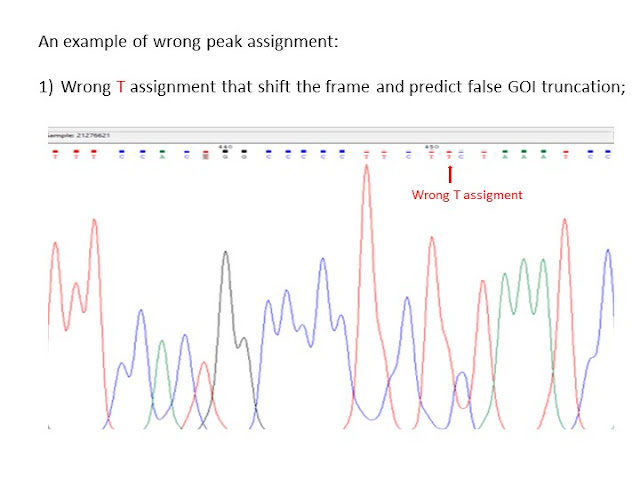
If the sequence show some point mutation or shift, do not  discard the clone but check carefully the chromatogram to see if the mutation/shift is real or is it due to a non-correct Chromas assignment that may happen in case of:
-  overlapping peaks:
- peak with low intensity;
- a sequence regions with multiple residues of the same species;
  
Other similar softwares:


Wednesday, September 14, 2022

ProteoCool Pills#19: Pay attention to reverse GOI insertion in TOPO cloning

The TOPO cloning technology (compared with PIPE and standard cloning in ProteoCool n° 1 is a simply and fast cloning approach able to accommodate a wide range of PCR insert sizes.

TOPO technology enables inserts with compatible ends to be readily joined to the vector in 5 minutes, without the need for additional ligation steps.

In my experience in some cases, incorrect Gene insertion (reverse) may happen in a certain %  (in my expereince 40-50%) of the E.coli clones obtained from TOPO reaction: 

This problem could be generally overcame just by screening and sequencing a large number of colonies (at least 4-6 for each clone)

However is important to pay attention to it during the PCR colony screening and the following sequence check:

For example, PCR colony screening  (see ProteoCool n° 4 for more detail about it)  performed with primers those anneal in the vector backborne will be not able to differenziate clones with reverse insertion from the good ones. 

Since the regions of the TOPO vectors located around (just before and after) the GOI insertion sites are quite similar (especially in the case that the GOI is cloned in a plasmid that do not codify for any C- or N- terminal tag)  is possible that the reverse insertion cpuld be not revealed by a not expert users

Example of cloning of a C-terminal His-tagger GOI in pcdna 3.4 TOPO for recombinant protein expression in mammalian cells (eg Expi293 or ExpiCHO)

In this case primers in addition to the GOI annealing sequence have to contain a flag carryng:

- KOZAC sequence before ATG start codon (Forward primer); 

- codons codyfing for the His tag followed by 1 or 2 Stop codons;

The final expected sequence (in the GOI is inserted in the correct direction) will be:

 TGACCTCCATAGAAGACACCGGGACCGATCCAGCCTCCGGACTCTAGAGGATCGAACCCTTgccaccATG-GOI-HIStag-STOP -STOP -AAGGGTTCGATCCCTACCGGTTAGTAATGAGTTTGATATCTCGACAATCAACCTCTGG

In yellow --> The region of pcdna3.4 before the insertion,

In Green --> The region of pcdna 3.4 after the insertion; 

and the respective Aminoacid traslation will be:

DLHRRHRDRSSLRTLEDRTLATM-GOI-HisTag**KGSIPTG***V*YLDNQPL 

 where * indicates STOP CODONS

On the contrary in case that the GOI is inserted in the opposite direction the final sequence will be:

ATTTTGTAATCCAGAGGTTGATTGTCGAGATATCAAACTCATTACTAACCGGTAGGGATCGAACCCTTgccaccATG-GOI-HIStag-STOP-STOP-GGGAGGGGGAAAGCGAAAGTCCCGGAAAGGAGCTGACAGGTGGTGGCAATGCCC

and the respective Aminoacid traslation will be:

FVIQRLIVEISNSLLTGRDRTLATM-GOI-HISTag**KGSIL*SPEAGSVPVSSMEVKTAWM

Performing an allingment you can easly see how the insert flanking regions are really similar

therefore during the sequence check, the sequence analisys cannot be restricted to the few bases close to the insert but need to be extended at least to 10-20bp before and 15-20bp after the insert. 

The risk of this kind of mistake is higher in vectors as pcdna3.4 those do not codify for any additional N- or C-terminal tag. In a vector those codify for a TAG, in case of reverse PCR insertion, you will found the  AA sequence of the TAG traslated in the opposite direction respect than the sequence of the insert.

In case you have some clone produced using TOPO cloning and do not show any expression,, i suggest to you,  before re-design the cloning strategy, to perform a double sequence check to be sure that your gene was inserted in the correct direction. 


Thursday, September 9, 2021

ProteoCool Pills#12: Rapid insertion/deletion/replacement of N-terminal signal peptide sequences using PIPE cloning

 In both eukaryotic and prokaryotic cells, all proteins are synthesized in cytoplasm. 

Proteins that are destined to enter into the secretory pathway are usually endowed with an N-terminal signal peptides (SPs, known also as N terminal leader sequences): the signal peptide those are short peptides and usually have a length of 16–30 amino acids.

After directing proteins to their specific locations, SPs are removed by signal peptidases

The presence or absence of the SPs allow to direct the expression of the protein in different cellular compartments:

- E.coli,  a protein with-out SPs will be directed in the cytoplasm, while the addiction of a SPs (as pelB, OmpA signal peptides) of the signal will  direct the protein into the periplasm;

-In Gram positive bacteria (as baccilus) and mammalian cells (as HEK293, CHO) the addiction of N terminal SPs direct the protein in the culture surnatant.

Since the accumulation of recombinant proteins in the cytoplasm may lead to the formation of inclusion bodies or protein degradation via proteases and the recombinant protein folding may also affected from the reducing conditions of the cell compartment (eg E.coli cytoplasm is strongly reducing and not compatible with S-S bond formation), the selection of the right Signal peptides play a decisive role in the industrial production of recombinant proteins.

It has been shown that using different homologous or heterologous signal peptides can affect the yields of recombinant proteins. Selecting a proper signal peptide to increase the secretion efficiency becomes a common methodology to optimize the production of recombinant protein

The availability of a simple cloning method to readily add, replace and modify a signal peptide sequences in an expression clone is therefore an essential tool to screen for the best protein/antibody productivity.

As already mentioned in the ProteoCool n°1: Cloning methods overview; The PIPE cloning is a nice method to manipulate expression vectors and perform mutagenesis, insertion and/or deletion or some vector regions.

With a single vector PCR is possible to insert dna fragments up to 80-100bp in any  vector region and therefore it can be applied also to the insertion of replacement of signal peptides in the vector of interest.


Using the PIPE cloning i  was able to insert pelB (MKYLLPTAAAGLLLLAAQPAMA)  and ompA (MKKTAIAIAVALAGFATVAQA) signal peptides in several pet15 clones for E.coli expression and other mammalian signal peptide in pcdna 3.4 clones for expression in Expi293 and Expi-CHO.

Of couse a similar approach could be performed to insert/delete or replace other short sequences as:

 - 6x His Tag
- Avi Tag
- Protease digestion sites (eg TEV, Eterokinase, Fatt.Xa)

or delete:
-N- or C- teminal region of your gene;
-N or C- terminal fusions (eg GFP, GST, MBP, etc)
----------------------------------------------------------------------------------------------------------------------------

PIPE cloning steps:

1) Vector amplification by PCR  (V-PCR)

2) PCR digestion with dpnI (to remove template vector background)

3) Trasfrom the vector into the MACH1 E.coli cells

4) Plasmid extraction from at least 4 colonies and DNA sequencing

-----------------------------------------------------------------------------------------------------------

                                            Tips to perform the Vector PCR:

PCR reaction:

My preferred DNA polymerases: Kapa Hifi (Roche) or Clone amp (Takara)

                       Theoretically you can use all high fidelity polimerases that do not add poly AA

DNA template --> less than 0.1ng/reaction 

(higher template amount may result in background colonies with the original template)

PCR volume --> 25ul/reaction are more than enough considering that for PIPE reaction 1-2ul are normally enough and PCR purification is not required.

PCR cycle:

Elongation time --> >1minute/kb also if the datasheet of the Taq suggest shorter extension time to exploit the 3-' --> 5' exonuclease activity that improve the formation of incomplete extension that are require for PIPE cloning.

Do not insert the final elongation time step.

---------------------------------------------------------------------------------------------------------------------------  

V-PCR trasfromation in E.coli cells (PIPE reaction)

PIPE cloning do not require PCR purification.

1)  Mix 2 ul of the V-PCR in 20ul of chemically competent MACH1 cells (thermo cod. )and incubate the mixture in ice 30 minutes;

N.B:  If the V-PCR band intensity is very high  (as in the reported picture)  yon can try also to dilute the plasmid 5 times and perform a second trasfromation with 2ul of the diluted plasmid in 20ul of cells because too much DNA can reduce the trasformation efficiency)

2) Incubate the cells 1' at 42°C

3) Transfer the cell in ice

4) Add 250ul of SOC or LB sterile media (with out antibiotic)

5) incubate the cells at 37°C - 180/600rpm (in a thermomixer or incubator shaker) 

6) Plate all the cells in LB-agar plates containing the proper selection antibiotic

(eg 100mg/l ampicillin for pet21 or pcdna 3.4,  50mg/l of kanamicin for pet24 clones)

7) incubate the plates O/N at 37°C

Primers design

Annealing regions à 18-26bp with an annealing temperature salt adjusted (calculated with Oligocalculator)  of 58-62°C if is possible)

Flanking regions –> length up to 60bp. Overlapping regions have to be between 15 and 20bp

Is not mandatory to add flanking regions in both forward and reverse primer to create the 16bp overlapping. It can be done also adding the flanking region in just 1 of the 2 primers. I choose one or the other solution on the basis of the length and Tm of the relative annealing regions.

----------------------------------------------------------------------------------------------------------------------------------------------------                                                                                      A great thanks to  

Roberto Petracca

an amazing supervisor who introduced me to the PIPE cloning

i was very lucky to shave with him many years in the Novartis Reseach centre in Siena 

References: 

Klock HE, Lesley SA. The Polymerase Incomplete Primer Extension (PIPE) method applied to high-throughput cloning and site-directed mutagenesis. Methods Mol Biol. 2009;498:91-103.


ProteoCool Pills #32: HemA, a powerfull selection marker for antibiotic free plasmid mantainance for recombinant protein expression in E.coli

Antibiotic resistance genes  (e.g Ampicillin , Kanamycin ) are the most commonly used markers for plamisd selection in DNA production and r...