Showing posts with label mab. Show all posts

Thursday, June 12, 2025

ProteoCool Pills #31: Modern Approaches to modulate the antibody effector function


Full length human IgG antibodies are composed of 2 regions, the N-terminal region of each chain corresponding to the Fab (fragment for antigen binding which contain the variable regions and the CDRs) and the C-terminal region corresponding to the Fc (crystallizable fragment)

Until some years ago the scientist focus mainly their efforts in the optimization of the Fab regions to develop antibodies with highest antigen affinity and lower immunogenicity.

In the last years, especially with the emergence of bispecific antibodies, e.g T-cell engagers there has been a growing interest in modifying the Fc region to modulate (enhance or remove) the antibody effector function.

The Fc region is able to bind with high affinity the Fc gamma receptors (FcγRs), and the neonatal Fc receptor (FcRn) expressed in the surface of the immune system cells (Reference)  by triggering  different effector functions such as antibody-dependent cell-mediated cytotoxicity (ADCC) and antibody-dependent cellular phagocytosis (ADCP) and complement- dependent cytotoxicity  (CDC) responses (Reference)

Among the four subclasses of human IgG (IgG1, IgG2, IgG3, IgG4), which differ in their constant regions, particularly in their hinges and CH2 domains, IgG1 has the highest FcγR-binding affinity, followed by IgG3, IgG2, and IgG4. As a result, different subclasses modulate in different way the different responses ADCC, ADCP and CDC.  (Reference)

In in the first generation of monoclonal antibodies the Fc activity was modulated by producing of the antibody in different Fc format on the basis of the antigen properties and the MoA (mechanism of action) that they would confer to the antibody: 

Blocking antibodies that do not have to activate the immune response where produced in the IgG4 format

For example Nivolumab which binds to the PD-1 receptor located on the cell membrane and inhibits its interaction with its ligands, PD-L1 and PD-L2. Since PD-1 is expressed on activated T cells, NK cells, regulatory T cells and B cells. Nivolumab has been desinged to block the interaction with the PD-L1 and PD-L2 and therefore releases immune cells from pathological immune suppression, but not activate the immune-response against these immune-cells otherwise we will obtain the unwanted depletion of effector cells inducing  serious adverse events..

On the contrary, mabs targeting antigen expressed in the surface of cancer cells (e.g Cetuximab targeting EGFR) were produced in igG1 form to activate the immune-response against those cells and induce cell clearance. In fact, it has been demonstrated in preclinical models and ex vivo studies that target-bound cetuximab IgG1 isotype mAbs stimulate natural killer (NK) cell–driven cytotoxicity against tumor cells coated in mAbs via the interaction of the constant region and the CD16 receptor on NK cells

More recently  igG1-Fc sequence engineering were largely exploited to further improve the modulation of Fc function:engineering as well as CHO cell line 

Fc Function enhancement:

It is well-known that this effector function is modulated by the N-linked glycosylation in the Fc region of the antibody (N297 of IgG1).


 In particular, absence of core fucose on the Fc N-glycan has been shown to increase IgG1 Fc binding affinity to the FcγRIIIa present on immune effector cells such as natural killer cells and lead to enhanced ADCC activity.

As I reported in the ProteoCool Pills #10, the simple supplementation of the culture media with  2F-Peracetyl-Fucose allow to produce low fucosilated monoclonal antibodies in mg scale to be used for R&D screening, on the other hand, several strategies (as the GlymaxX® and POTELLIGENT®  have been developed to build stable clones for large scale production based of afucosylated antibodies with improved therapeutic potency.

Since those cell lines may show some drawbacks in terms of growth rate and mab productivity at the same time other scientists focus also in the development of igG1 mutant (eg S239D/1332E  named SDIE and G236A/S239D/A330L/I332E named GASDALIE – US patentUS20230057150A1  those similarly to afucosilated mab show improved affinity for the FcγRs even in produced in the standard CHO cell lines.

In the past I had the opportunity to do some trials comparing the ADCC activity (using the Promega ADCC assay, both F and V versions) with using the igG1 WT afucosilated and igG1 SDIE mutant (produced with standard fucosilation) for the same clone and interestingly  (data not shown) it seems that their effect could be combined at least from the results of this in vitro assay. 

B) Fc Function silencing:

The most recent alternative to the use of IgG1, which allow to point mutations in the linker region between hinge and Fc domains were described to reduce or fully abrogate Fcγ binding affinity and downstream effector cell activation

In the following Table are reported some of the most known and used mutations:
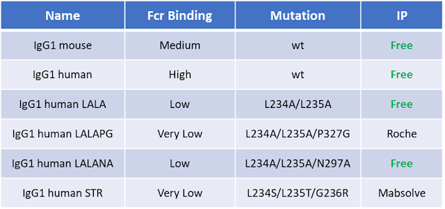

From literature the STR (recently developed by MAbSolve seems to be the only mutation that completely abolish all the ADCC, ADCP and CDC activities (Reference) and  it seems that STR do not alter the antibody developability profile.

Even in this case I had the opportunity to do some R&D trials comparing the igG1 human wild type, LALAPG and STR mutants and I was not able to was not able to reveal meaningful differences between LALAPG and STR with the Promega ADCC assay (complete abolishment in both cases – data not shown) but this can be due to the fact that as shown in seems that the advantage of STR vs LALAPG is in the lower affinity of STR for the FcγRI which is probably more involved in trigger the ADCP than ADCC response or that the sensitivity of those kind of assay is not enough to detect so small differences (see Table3 the authors do not reveal any differences even in ACDP assays

I also performed antibody aggregation propensity detected performing SEC on 10mg/ml samples subjected to different incubations (4°C, 37°C, Freeze/Thaw) and in this case the LALAPG seems to be slightly better (data not show)

However, this post does not would provide any ranking or evaluation of the different mutants but just inform you about the possibility to test them to achieve the wanted MoA for your new monoclonal antibody.


Sunday, August 4, 2024

usefull links #1

i would like with share the folliwng 3 links about usefull on line tool for the scientist working with recombinant monoclonal antibodies: 

 1) ABRSA is a simple and useful tool for antibody numberimg and cdr identification 

 2) PLABDAB is a repository containing over 150,000 paired antibody sequences derived from patents and academic paper. it is vero usefull to underand if a mab sequence is patented or not. 

 3) TAP is an on line tool to idenify criticism that cam affect mab developabilit

 #mab #onlinetools #science #cdr #antibodynumbering

Monday, June 5, 2023

ProteoCool Pills#27: DSF an useful tool for Monoclonal antibodies selection, optimization and developabiltiy assesment


DSF 

(Differential scanning fluorimetry previously named also Thermofluor)

is a very simple tecniques that exploit the ability of a fluorescente probe,  typically either SYPRO Orange or 1-anilino-8-naphthalenesulfonate (ANS) – that is quenched in an aqueous environment but becomes strongly fluorescent when bound to exposed hydrophobic groups of a protein. Therefore, the thermal unfolding of a protein of interest in the presence of such a dye, can be followed spectrophotometrically.

DSF is a 96 well based assay that could be performed on a RT-PCR thermocycler, it is inexpensive,  fast, and require relatively little sample. All these advantages have made this approach attractive for screening applications in drug discovery  and also for protein stability formulation

In the ProteoCool n°24 in suggest as in my opinion the DSF may become an essential assay todetermine lot to lot concistenciy in QC department of company producing recombinant proteins (e.g recombinant vaccine and/or enzymes)

In this post i would like to share with you 2 examples that show as DSF may play an important role also in antibody screening, optimization and developability assesment. 

DSF thanks to its excellent throughput and minimal material consumption (about 50ul of antibody at 0,3 mg/ml for each point) allow to readily compare the thermal stability of:

- different antibodies in the same buffer:

-  different  Fab mutants or Fc mutants in the same buffer;

-  different  antibody formulations; (optimize pH, salt concentration, excipients)


Example 1: 

Comparision of thermal stability of different mabs 

Full length human IgG1-Clk mabs  in PBS may show 1 or 2 separate transitions on the basis of the Fab stability. 

Mabs with lower Fab stability show in general a single transition while mabs with high stability show 2 separate transitions, 1 at about 69°C for the Fc and 1 at higher temperature for the Fab.


Example 2:

Comparision of thermal stability of different Fc mutants

In recent years the enhancement or elimination of the Fc-effector function has led to a growing interest in Fc-engineering, to give antibodies specific mechanisms of action and therapeutic properties.  

Even if several Fc modification, as:

-   LALANA, LALAPG, LALAGR mutations to remove effector function;

SDIE, GAALIE mutations as well as afucosilation to enanche the effector function

 were already reported, few data about developability of those solutions are still available and there is a lot work  to do to select an enanched Fc combining the best effector function and good manufacturability, stability and pharmacokinetics.

Those are just 2 examples that show as in short time and using a limited amount od samples, thanks to DSF we can obtain  essential informations to guide our decision in antibody during the screening, desing, formulation phases.

Of course, thermal stability itself do not Guarantee that the selected antibody has good developability profile but  some of the following other tecniques as:

- DLS;

- PEG solubility;

- Self-interaction determined by BLI;

those provide informations about antibody  aggregation propensity 

- Hydrofobic interaction chromatografy (HIC)

those provide informations about antibody hydrofobicity

have to be peformed to complement the DSF data. 


A great thanks  to

 Luca Sorrentino


Sunday, October 9, 2022

ProteoCool Pills#20: Micro PEG solubility screening, a simple Tool for Biologics Design and Formulation Development.

Adequate protein solubility is an important prerequisite for development, manufacture, and administration of biotherapeutic drug candidates, especially for high-concentration protein formulations. 

For example, in monoclonal antibody discovery, early identification of monoclonal antibody candidates whose development, as high concentration (≥100 mg/mL) drug products, could prove challenging, due to self-interaction that may induce high viscosity, can help define strategies for candidate engineering and selection.

If in theory, Rheology measurements are an effective means for characterizing therapeutic protein/antibody solutions, practically, the conventional measurements are hindered by the limited amount of material, especially during early development, when it is necessary to screen and compare several different molecules over a wide range of conditions (e.g different pH, additives, concentration)

Therefore alternative techniques able to provide hints about aggregation propensity and solubility using a smaller sample volume are essential to compare and select the best candidates in the early development and reduce the risk of move forward an candidate with high developability risk. 

Dynamic light Scattering (DLS) is probably the most used technique for this purpose since it allows to:

1) Characterize the sample intermolecular interactions (attractive or repulsive?) comparing how the diffusion coefficient (Dt) is affected by concentration since: 

In an ideal dilute solution, the diffusion coefficient (Dt) measured by DLS is not dependent on solute concentration. As concentration increases, the solution becomes less ideal. 

           Dt=D0(1+kD*C)

Attractive interactions (kD < 0) cause an apparent decrease in Dt and an apparent increase in Rh, while repulsive interactions (kD > 0) cause an apparent increase in Dt and an apparent decrease in Rh 

Therefore, decrease of Dt in function of the concentration, indicating the presence of repulsive intermolecular interactions while increase of Dt in function of the concentration, indicate presence of attractive interactions (sample more prone to aggregation)

2) Perform viscosity assessment by Microrheology: Using polystyrene beads with known values of R allows for the determination of the viscosity of the protein solution that the beads are suspended in. The size of the beads is larger than that of the protein molecules, and thus the DLS signals can easily be separated.

Bilayer interferometry (BLI) was also recently proposed as an alternative to DLS to assess protein self-interaction. (Sun et.al mabs 2013Domnowsky et. al International Journal of Pharmaceutics 2020

All those methods are fast and require a small amount of material but they require specific and expensive instrumentations those are not present in all the laboratories.

In this post, I would like to introduce you a simple method that could be done in every laboratory (since it requires the presence of a centrifuge for plates and nanodrop UV spectrophotometer or similar) based on PEG precipitation, a previously established method for determining the relative apparent solubility of adapted for screening in small scale and which is reported to correlate with the Kd values obtained by DLS (Scannell et.al, Pharm Res 2021

This method, adapted in 96 well plate allow to compare monoclonal antibody (mAb) candidates also if only limited quantities (eg. 1 mg) are available. 

Protocol 

(for A280nm reading with Nanodrop or similar)

(adapted from Toprani et.al J Pharm Sci. 2016

Day1 (afternoon)

1) 25ul of monoclonal antibody at 1mg/ml mixed with 25ul of PEG10K solutions at different concentrations (from 32% to 8%) in a 96well V-bottom plate

                           Example of a plate assembled to test 6 different mabs in duplicate: 

2) The plate was covered by aluminum foil and incubated O/N at 4°C

Day2

3) The plate was centrifuged 1h at 3200g at 4°C; 

4)10ul of surnatant were carefully transferred in a 96 well PCR plate (using a multichannel pipette). V bottom plates are preferable since the form of the well reduce the probability to resuspend the precipitate during the surnatant pick-up;

5) Amount of mab present in the surnatant was quantified by measuring the A(280nm) by NanoDrop Spectrophotometer;

6) Relative soluble fraction is calculated and plotted as function of PEG concentration;

Example of results

Example 1

 Comparision of  PEG solubility for 3 different mabs  (human igG1-CLk) in PBS buffer


Example 2

Comparision of PEG solubility for 2 different mabs in 2 different buffers (different pH)


The main limit of this protocoll is represented by the throughput, since sample reading by nanodrop allow to scale down the protocol and reduce a lot the protein amount but it is not very fast. 

6 mabs in duplicate --> 96 well --> more than 1h at nanodrop

For high number of samples, you can run a modified verision of the protocol, based on A280 deterination using 96well half volume UV clear plates (Greiner cod. 675801) in a multiplate reader. 

Since, in multiwell reader, the A280nm value change in fuction of both, concentration and optical path (that is function of the sample volume)  in this case to obtain a good sensitivitty the reaction volume (step1) were doubled  (50ul of mab 1mg/l + 50ul of PEG solution) and after centrifugation 70ul of the surnatant were tranfered to the  UV-clear 96 plate for the A280 determination with the multiplate reader. 

6 mabs in duplicate --> 96 well --> 1-2 minutes

Example 3

Comparision of PEG solubility for a wild tipe mab vs some mutant in PBS 

A(280nm) measure with Biotek- cytation5 multiplate reader

Microplate reader vs nanodrop:

Pros:
 Throughput (A plate could be acquired in few minutes)

Cons: 
Double amout of material required: (0,4 mg vs 0,2mg of mab each coloum)
Cost of the plates (about 8 euro/plates

Materials: 

- PEG 10K (Alfa Aesar cod. B21955)
- 96 well V-bottom plates (Costar cod. 3897)
- 96 UV-clear half volume (Greiner cod. 675801 cost ~ 8 euro/plate)

Other references: 

https://www.americanlaboratory.com/media/20/Document/DLS-Microrheology.pdf


Wednesday, July 27, 2022

ProteoCool Pills#18: MabSelect@ a cheaper alternative to proteinA sepharose FF resin for small scale gravity flow purification of recombinant mab

Affinity chromatography which is based on the interaction the Fc region of the mAb molecule with  specifi bacterial proteins as proteinA, proteinG or protein L immobilized on the resin is generally used for the isolation of antibodies from culture surnantants of the cell lines (eg CHO) used for their recombinant production.

The binding specificity and strength of protein A, protein G and protein L is not equally strong for all immunoglobulins and, in the case of IgG, not equal for all isotypes (Link1, Link2)

A generally stronger binding to the Fc region is observed by protein G, however higher binding strength however does not automatically result in better results since also the presence of impurities may influence the binding capacity.

In both, protein A and protein G affinity chromatography, the elution is carried out using a low pH buffer (eg glicine pH 2,7 for proteinG, and citrate pH3,0 for protein A). Generally in protein G chromatography, a stronger eluent is required to elute the  captured antibody from the coloumn.

Thus, protein A chromatography is preferred over protein G since lower levels of impurities are generally obtained and it currently represent the gold standard in mAb pruficiation. 

In general an efficient protein A resin should have: 

- High dynamic binding capacity (able to bind large amounts of mAbs in a short time) which allow high flow-rate without losing mAbs in the flow-through

-  High stability of the resin under regeneration with sodium hydroxide. The number of cycles you can run with the same resin has a huge economic impact;

Often, in the preliminary phases of the pre-clinical research, to identify the best mabs, scientists have to a large panel of different mabs in small amount (from ug to mgs). In absence of robotic platforms dedicated to mab purification, the gravity flow purification may represent a simple and powerful alternative.   

Gravity flow purification require a resin with high porosity, rigidity and low backpressure to avoid resin clogging and guarantee reasonable flow-rate and purification timelines.

The Citivya rProteinA Fast flow resins  (90uM of particle diameter), which represent the gold standard for the gravity flow purification of recombinant monoclonal antibodies is very expensive  (more than 80euro/ml). 

If it is true that this resin could be cleaned and re-generated several time and that thanks to its high binding capability (>35mg/ml)  generally small volumes (100-500ul/sample) of resin are enough to purify mgs of mab samples requited in the preliminary mab screening phase i however its cost may have an high impact expecially in academic laboratories and the identification of an alternative resin with similar performances but low cost is preferable.

In this post i would like to share with you some enocuraging results that i have recently obtained with gravity flow purification by replacing the rprotA FF with the cheaper Mabselect resin which is reported to have 

- similar binding capability  (30mg/ml of Mabselect vs 35mg/ml of rproA FF)

and

- similar particle size  (85um of Mabselect vs 90um of rproA FF)

but it is at least 5 time cheaper (25ml of mabselect cost = 5ml of rprotA resin)


Mab Select resin tested in the 2 following  formats

format 1 (small)  --> 75ul of resin in a Poliprep coloum (Biorad cod. #731-550)

format2 (medium) --> 750ul of resin in a PD-10 empty coloumn  (GE cod. 17-0851-01)

showed flow rate very close to the FF (as you can see in the following videos: protA FF on the left, MAbselect on the right) 

Video 1: Equilibration with buffer


                                                 Video 2: Expi-CHO surnatant loading


and similar results in terms of binding capacity and final mab purity level

In the following picture, 2 examples of purification performed with the MabSelect resin

In both cases the following buffers were used: 


- Tris 2M pH=8 (1ml for 20ml culture) to correct the surnatant pH before coloumn loading;

- Equilibration and washing buffer; Tris 25mM, NaCl 25mM pH 7,2  buffer;

- Elution buffer: 300ul di Citrate 100mM NaCl 60mM pH=3;

- Tris 1M pH=9 (30ul for small size; 300ul for medium size) to neutralize the acid pH after elution


Of course there are many other interesting proteinA resins cheaper than the rProtA FF ND produded from suppliers different from Cytiva as  Tosoh. Biorad,  Thermo that could be also tested.

Personally, i selected the Mabselect since was the one with the particle size more close to the FF and i suspect that this detail can be essential to guarantee a good flow rare  in gravity flow purification. 

Friday, April 29, 2022

ProteoCool Pills#16: Loading small sample volumes on AKTA instruments

AKTA instruments (as AKTA-FPLC, AKTA-PRIME, AKTA-Purifiers, AKTA-Go, AKTA-pure) represent in the last 30 years the point of reference for protein/antibody purification in both research, development and pharmaceutical production departments since they are robust, easy to use and provided with a software (the Unicorn)  which is a reall uses friendly software.

On the contrary, their application for protein/antibody analytics characterization is mainly limited to some specific columns and approaches (eg SEC chromatography with the Superdex 10/30 or 5/15 coloumns) in those laboratories that do not have budget dedicate to acquire an analitical HPLC. 

 In those case the loading and injection of a small sample volume in reproducible way it a prerequisite.

How we can inject small volumes in an FPLC or other AKTA instruments? 

For volumes between 200-300µl to 1ml,  the standard injection port could be used with an 1ml insulin plastic syringe (with out needle)


For volumes <250µl ,  the standard injection port need to be replaced with a injection filling port designed for small volumes and use an more precise Hamilton syringe (250µl or 100µl volume) for sample injection.  

Until some years ago (as I already wrote to answer to a question in Research gate in 2019) https://www.researchgate.net/post/Loading-small-sample-volumes-on-FPLC), Citiva bioscience (previously GE bioscience) supplied for small sample injection a metal injection filling port (CITIVA cod. 19768701) supplied with a plastic syringe stand suitable for injection of small volumes using Hamilton syringes

Recently those Injection fill port was discontinued and replaced by a plastic filling post (Fill Port, INV-907 cod.18112766)

Also if this filling port, is not supplied with any syringe stand, it could be used directly with an hamilton syringe, as you can see from the following imagine:


Of course to inject so small volumes you need to select also the appropriate:

  •          Injection loop (with a small volume)
  •         Syringes (with BLUNT tips:

Suggested:

50-100ul sample:  Hamilton 1710N volume 100 μL, needle size 22s ga, needle L 51 mm; Sigma-Aldrich cod. 28634-U cost ~100€;

250-100ul samples: Hamilton® 1725N, volume 250 μL, needle size 22 ga (blunt tip), needle L 51 mm; Sigma-Aldrich cod. 28636-U cost ~80€;


Wednesday, July 14, 2021

ProteoCool Pills#10: Use of 2F-Peracetyl-Fucose to produce low fucosilated monoclonal antibodies with Expi293 and ExpiCHO cell lines

Therapeutic monoclonal antibodies are the fastest growing class of therapeutics for the treatment of various cancers and inflammatory disorders. The Fc region of human IgG antibodies interacts with multiple Fcγ receptor (FcγR) and complement proteins and mediates immune effector functions, which are important for many therapeutic applications, e.g. elimination of targeted cells via antibody-dependent cellular-cytotoxicity (ADCC), -phagocytosis (ADCP) or complement-dependent cytotoxicity (CDC).

To date, therapeutic IgG antibodies (either approved or in clinical development) belong to the IgG1, IgG2 or IgG4 subclasses. Each IgG isotype has a distinct binding affinity to the various FcγRs, which are expressed differently on immune cells. 

In cancer immunotherapy, some IgG1 antibodies (eg ipilimumab) rely on the Fc-mediated immune effector function, dependent cellular cytotoxicity (ADCC), as the major mode of action to deplete tumor cells.

It is well-known that this effector function is modulated by the N-linked glycosylation (N297) in the Fc region of the antibody.

In fact, if the N297A mutation  is able to reduced FcγR-binding and abolish ADCC activity on the other hand,  the absence of core fucose on the Fc N-glycan has been shown to increase IgG1 Fc binding affinity to the FcγRIIIa present on immune effector cells such as natural killer cells and lead to enhanced ADCC activity.


Various strategies have focused on producing afucosylated antibodies to improve therapeutic efficacy. While engineering of the cell lines (eg FUT8 deletion: @Potelligent or @GlymaxX Technologies) are required for scale up production of recombinant afucosilated mabs, for small lab scale for production of mgs, the supplementation of 2F-Peracetyl-Fucose is a simple and efficient solution to produce “low fucosilated mabs” to be used in functional in vivo and in vitro assay.

Media supplementation with 200 µM of  2F-Peracetyl-Fucose allow to produce low fucosilated mabs with transient transfection in Expi293 or ExpiCHO cells) just before cell transfection (as reported on Chakraborty et.al for the Expi293 cells) allow to produce a mab with low fucosilation level (as reported in Fig1) and enhanced ADCC activity.

n.b: The results of ELISA assay on mabs produced from Expi293 and ExpiCHO seem to suggest that the ExpiCHO show a decreased fucosilation level respect than Expi293 also in absence of the 2F paracetyl fucose. 

Preparation of 2F-Peracetyl-Fucose stock solution:

2F-Peracetyl-Fucose is supplied as powder (10mg). It can be directly resuspended in the glass bottle with 1mM DMSO to obtain a 34.2mM stock solution (171X) that could be stored at -20°C.

584µl of 34.2mM of 2F-Peracetyl-Fucose were added to 100 ml of cells just before the transient transfection with Expifectamines

Prons:

Very Simple!!

Concerns:

Expensive: Cost of  2F-Peracetyl-Fucose: ~ 1000euro/liter of transfection make this approach feasible only for small lab scale productions


ProteoCool Pills #32: HemA, a powerfull selection marker for antibiotic free plasmid mantainance for recombinant protein expression in E.coli

Antibiotic resistance genes  (e.g Ampicillin , Kanamycin ) are the most commonly used markers for plamisd selection in DNA production and r...